N-localizer
| N-localizer | |
|---|---|
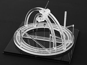 Three N-localizers attached to a stereotactic frame.[1] | |
| Specialty | neurosurgery, radiation oncology |
| Intervention | stereotactic surgery, radiosurgery |
| Inventor(s) | Russell A. Brown[2] |
[edit on Wikidata] | |
The N-localizer[3] is a device that enables guidance of stereotactic surgery or radiosurgery using tomographic images that are obtained via computed tomography (CT),[4] magnetic resonance imaging (MRI),[5] or positron emission tomography (PET).[6] The N-localizer comprises a diagonal rod that spans two vertical rods to form an N-shape (Figure 1) and permits calculation of the point where a tomographic image plane intersects the diagonal rod. Attaching three N-localizers to a stereotactic instrument allows calculation of three points where a tomographic image plane intersects three diagonal rods (Figure 2). These points determine the spatial orientation of the tomographic image plane relative to the stereotactic frame.[7]
The N-localizer is integrated with the Brown-Roberts-Wells (BRW),[8] Kelly-Goerss,[9] Leksell,[10] Cosman-Roberts-Wells (CRW),[11] Micromar-ETM03B, FiMe-BlueFrame, Macom, and Adeor-Zeppelin[12] stereotactic frames and with the Gamma Knife radiosurgery system.[13]
An alternative to the N-localizer is the Sturm-Pastyr localizer that comprises three rods wherein two diagonal rods form a V-shape and a third, vertical rod is positioned midway between the two diagonal rods (Figure 3).[14] The Sturm-Pastyr localizer is integrated with the Riechert-Mundinger and Zamorano-Dujovny stereotactic frames.[15]
Compared to the N-localizer, the Sturm-Pastyr localizer is less accurate and necessitates more elaborate calculations to determine the spatial orientation of the tomographic image plane relative to the stereotactic frame.[16] In contrast to the N-localizer that does not require specification of the pixel size in a tomographic image,[17] the Sturm-Pastyr localizer requires precise specification of the pixel size.[18]
Research conducted four decades after the introduction of the N-localizer[19] and Sturm-Pastyr localizer[20] has revealed computational techniques that improve the accuracy of both localizers.
Figures
-
 Figure 1. Depiction of the N-localizer and its intersection with the tomographic image plane. (A) Side view of the N-localizer. The tomographic image plane intersects two vertical rods and one diagonal rod. (B) Tomographic image. The intersection of the tomographic image plane with the N-localizer creates two fiducial circles and one fiducial ellipse. The relative spacing between the ellipse and the two circles varies with the height at which the tomographic image plane intersects the diagonal rod.
 -
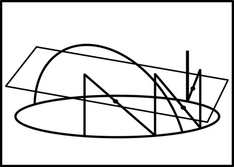 Figure 2. Depiction of three N-localizers and their intersection with the tomographic image plane. The quadrilateral represents the tomographic image plane. The oval and the arch represent the stereotactic instrument. The vertical and diagonal lines attached to the oval represent three N-localizers. The three points where the tomographic image plane intersects the diagonal rods are depicted by the dots. These points of intersection determine the spatial orientation of the tomographic image plane relative to the stereotactic frame.
 -
 Figure 3. Depiction of the Sturm-Pastyr localizer and its intersection with the tomographic image plane. (A) Side view of the Sturm-Pastyr localizer. The tomographic image plane intersects two diagonal rods and one vertical rod. (B) Tomographic image. The intersection of the tomographic image plane with the Sturm-Pastyr localizer creates two fiducial ellipses and one fiducial circle. The relative spacing between the circle and the two ellipses varies with the height at which the tomographic image plane intersects the vertical rod.


References
- ^ Arle, J (2009). "Development of a Classic: The Todd-Wells Apparatus, the BRW, and the CRW Stereotactic Frames". In Lozano, AM; Gildenberg, PL; Tasker, RR (eds.). Textbook of Stereotactic and Functional Neurosurgery. Berlin: Springer-Verlag. pp. 456–460. doi:10.1007/978-3-540-69960-6. ISBN 978-3-540-69959-0. S2CID 58803140.
- ^ "System Using Computed Tomography as for Selective Body Treatment". U.S. Patent 4608977. 1986.
- ^ Galloway, RL Jr. (2015). "Introduction and Historical Perspectives on Image-Guided Surgery". In Golby, AJ (ed.). Image-Guided Neurosurgery. Amsterdam: Elsevier. pp. 2–4. doi:10.1016/B978-0-12-800870-6.00001-7. ISBN 978-0-12-800870-6.
- ^ Thomas DG, Anderson RE, du Boulay GH (1984). "CT-guided stereotactic neurosurgery: experience in 24 cases with a new stereotactic system". Journal of Neurology, Neurosurgery & Psychiatry. 47 (1): 9–16. doi:10.1136/jnnp.47.1.9. PMC 1027634. PMID 6363629.
- ^ Heilbrun MP, Sunderland PM, McDonald PR, Wells TH Jr, Cosman E, Ganz E (1987). "Brown-Roberts-Wells stereotactic frame modifications to accomplish magnetic resonance imaging guidance in three planes". Applied Neurophysiology. 50 (1–6): 143–152. doi:10.1159/000100700. PMID 3329837.
- ^ Maciunas RJ, Kessler RM, Maurer C, Mandava V, Watt G, Smith G (1992). "Positron emission tomography imaging-directed stereotactic neurosurgery". Stereotactic and Functional Neurosurgery. 58 (1–4): 134–140. doi:10.1159/000098986. PMID 1439330.
- ^ Gildenberg, PL; Krauss, JK (2009). "History of Stereotactic Surgery". In Lozano, AM; Gildenberg, PL; Tasker, RR (eds.). Textbook of Stereotactic and Functional Neurosurgery. Berlin: Springer-Verlag. p. 23. doi:10.1007/978-3-540-69960-6. ISBN 978-3-540-69959-0. S2CID 58803140.
- ^ Heilbrun MP, Roberts TS, Apuzzo ML, Wells TH, Sabshin JK (1983). "Preliminary experience with Brown-Roberts-Wells (BRW) computerized tomography stereotaxic guidance system". Journal of Neurosurgery. 59 (2): 217–222. doi:10.3171/jns.1983.59.2.0217. PMID 6345727.
- ^ Goerss S, Kelly PJ, Kall B, Alker GJ Jr (1982). "A computed tomography stereotactic adaptation system". Neurosurgery. 10 (3): 375–379. doi:10.1227/00006123-198203000-00014. PMID 7041006.
- ^ Leksell L, Leksell D, Schwebel J (1985). "Stereotaxis and nuclear magnetic resonance". Journal of Neurology, Neurosurgery & Psychiatry. 48 (1): 14–18. doi:10.1136/jnnp.48.1.14. PMC 1028176. PMID 3882889.
- ^ Couldwell WT, Apuzzo ML (1990). "Initial experience related to the Cosman-Roberts-Wells stereotactic instrument. Technical note". Journal of Neurosurgery. 72 (1): 145–8. doi:10.3171/jns.1990.72.1.0145. PMID 2403588. S2CID 1363168.
- ^ Sedrak M, Alaminos-Bouza AL, Srivastava S (2020). "Coordinate systems for navigating stereotactic space: how not to get lost". Cureus. 12 (6): e8578. doi:10.7759/cureus.8578. PMC 7358954. PMID 32670714.
- ^ Tse, VCK; Kalani, MYS; Adler, JR (2015). "Techniques of Stereotactic Localization". In Chin, LS; Regine, WF (eds.). Principles and Practice of Stereotactic Radiosurgery. New York: Springer. pp. 25–32. doi:10.1007/978-1-4614-8363-2. ISBN 978-1-4614-8362-5.
- ^ Sturm V, Pastyr O, Schlegel W, Scharfenberg H, Zabel HJ, Netzeband G, Schabbert S, Berberich W (1983). "Stereotactic computer tomography with a modified Riechert-Mundinger device as the basis for integrated stereotactic neuroradiological investigations". Acta Neurochirurgica. 68 (1–2): 11–17. doi:10.1007/BF01406197. PMID 6344559. S2CID 38864553.
- ^ Krauss, JK (2009). "The Riechert/Mundinger Stereotactic Apparatus". In Lozano, AM; Gildenberg, PL; Tasker, RR (eds.). Textbook of Stereotactic and Functional Neurosurgery. Berlin: Springer-Verlag. pp. 487–493. doi:10.1007/978-3-540-69960-6. ISBN 978-3-540-69959-0. S2CID 58803140.
- ^ Alaminos-Bouza AL, Brown RA (2020). "Comparative accuracies of the N-localizer and Sturm-Pastyr localizer in the presence of image noise". Cureus. 12 (7): e9137. doi:10.7759/cureus.9137. PMC 7364427. PMID 32685325.
- ^ Weaver K, Smith V, Lewis JD, Lulu B, Barnett CM, Leibel SA, Gutin P, Larson D, Phillips T (1990). "A CT-based computerized treatment planning system for I-125 stereotactic brain implants". International Journal of Radiation Oncology, Biology, Physics. 18 (2): 445–454. doi:10.1016/0360-3016(90)90114-Y. PMID 2406230.
- ^ Dai J, Zhu Y, Qu H, Hu Y (2001). "An algorithm for stereotactic localization by computed tomography or magnetic resonance imaging". Physics in Medicine and Biology. 46 (1): N1–N7. doi:10.1088/0031-9155/46/1/401. PMID 11197682. S2CID 9196917.
- ^ Sedrak M, Alaminos-Bouza AL, Bruna A, Brown RA (2021). "Monte Carlo simulation of errors for N-localizer systems in stereotactic neurosurgery: novel proposals for improvements". Cureus. 13 (2): e13393. doi:10.7759/cureus.13393. PMC 7977485. PMID 33758694.
- ^ Alaminos-Bouza AL, Brown RA (2021). "Improved accuracy for the Sturm-Pastyr localizer in the presence of image noise". Cureus. 13 (9): e17905. doi:10.7759/cureus.17905. PMC 8509111. PMID 34660100.
Further reading
- Saleh, H; Kassas, B (2014). "Developing Stereotactic Frames for Cranial Treatment". In Benedict, SH; Schlesinger, DJ; Goetsche, SJ; Kavanagh, BD (eds.). Stereotactic Radiosurgery and Stereotactic Body Radiation Therapy. Boca Raton: CRC Press. pp. 156–159. doi:10.1201/b16776. ISBN 978-1-4398-4198-3. S2CID 58555632.


