Extensor pollicis brevis muscle
| Extensor pollicis brevis muscle | |
|---|---|
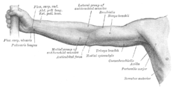 Front of right upper extremity. (Extensor pollicis brevis labeled at upper left.) | |
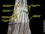
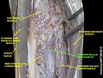
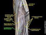
 Posterior surface of the forearm. Deep muscles. (Extensor pollicis brevis visible at left.) | |
| Details | |
| Origin | Radius and the Interosseous membrane |
| Insertion | Thumb, proximal phalanx |
| Artery | Posterior interosseous artery |
| Nerve | Posterior interosseous nerve |
| Actions | Extension of thumb at metacarpophalangeal joint |
| Antagonist | Flexor pollicis longus muscle, flexor pollicis brevis muscle |
| Identifiers | |
| Latin | musculus extensor pollicis brevis |
| TA98 | A04.6.02.050 |
| TA2 | 2518 |
| FMA | 38518 |
| Anatomical terms of muscle [edit on Wikidata] | |
In human anatomy, the extensor pollicis brevis (EPB) is a skeletal muscle on the dorsal side of the forearm. It lies on the medial side of, and is closely connected with, the abductor pollicis longus. The extensor pollicis brevis belongs to the deep group of the posterior fascial compartment of the forearm. It is a part of the lateral border of the anatomical snuffbox.
Structure
The extensor pollicis brevis arises from the ulna distal to the abductor pollicis longus, from the interosseous membrane, and from the dorsal surface of the radius.[1]
Its direction is similar to that of the abductor pollicis longus, its tendon passing the same groove on the lateral side of the lower end of the radius, to be inserted into the base of the first phalanx of the thumb.
Variation
Absence; fusion of tendon with that of the extensor pollicis longus or abductor pollicis longus muscle.
Function
In a close relationship to the abductor pollicis longus, the extensor pollicis brevis both extends and abducts the thumb[1] at the carpometacarpal and metacarpophalangeal joints.[2]
Additional images
-

-
 Bones of left forearm. Posterior aspect.
 -
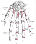 Bones of the left hand. Dorsal surface.
 -
 Tendons of forefinger and vincula tendina.
 -
 Transverse section across distal ends of radius and ulna.
 -
 Transverse section across the wrist and digits.
 -
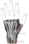 The mucous sheaths of the tendons on the back of the wrist.
 -
 The radial and ulnar arteries.
 -
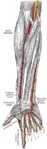
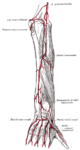 Arteries of the back of the forearm and hand.
 -
 Extensor carpi radialis brevis muscle
 -
 Extensor pollicis brevis muscle
 -
 Extensor pollicis brevis muscle
 -
 Extensor pollicis brevis muscle
 -
 Extensor pollicis brevis muscle
 -
 Extensor pollicis brevis muscle
 -
 Extensor pollicis brevis muscle
 -
 Extensor pollicis brevis muscle
 -
 Muscle of the hand. Posterior view.


References
![]() This article incorporates text in the public domain from page 455 of the 20th edition of Gray's Anatomy (1918)
This article incorporates text in the public domain from page 455 of the 20th edition of Gray's Anatomy (1918)
- ^ a b Platzer 2004, p. 168
- ^ "Thumb Articulations". ExRx.net.
Sources
- Platzer, Werner (2004). Color Atlas of Human Anatomy, Vol. 1: Locomotor System (5th ed.). Thieme. ISBN 3-13-533305-1.
External links
Wikimedia Commons has media related to Extensor pollicis brevis muscle.
- PTCentral
- v
- t
- e
| fascia: | |
|---|---|
(compartments)
| anterior | |
|---|---|
| posterior | |
| fascia |
|
| other |
(compartments)
| anterior |
| ||||
|---|---|---|---|---|---|
| posterior |
| ||||
| fascia |
| ||||
| other |
| lateral volar | |||||
|---|---|---|---|---|---|
| medial volar |
| ||||
| intermediate | |||||
| fascia |
|
Portal:
 Anatomy

| Authority control databases |
|
|---|


